Prepare to delve into the fascinating world of angiomas, benign tumors composed of blood vessels that can appear in various forms and locations throughout the body. From their captivating clinical presentations to the diverse treatment options available, this comprehensive guide will illuminate the complexities of angiomas.
Unravel the mysteries of these enigmatic growths as we explore their causes, risk factors, and potential complications. Discover the latest advancements in diagnosis and treatment, empowering you with the knowledge to navigate the challenges and triumphs associated with angiomas.
Definition of Angioma
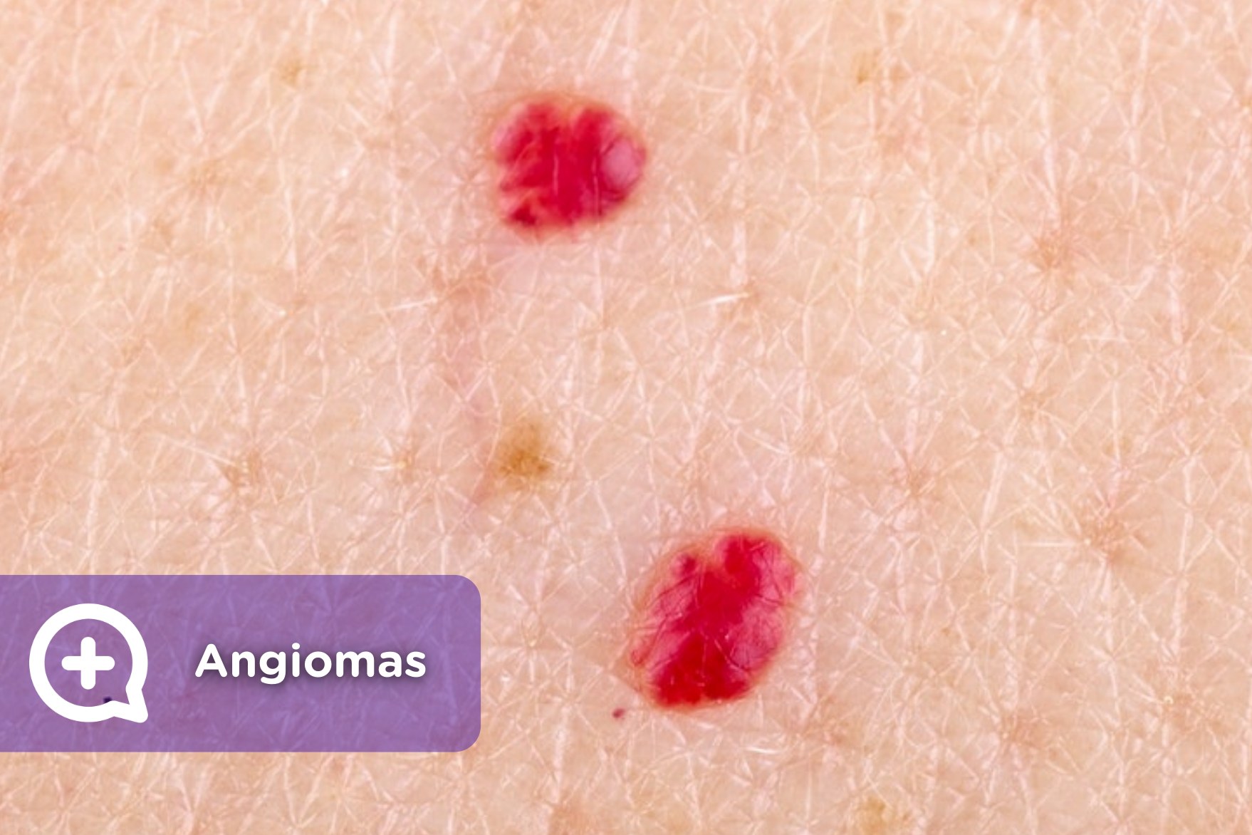
An angioma is a non-cancerous growth of blood vessels that can occur anywhere in the body. They are usually bright red or purple in color and can range in size from a few millimeters to several centimeters.
Angiomas are caused by an overgrowth of blood vessels. The exact cause of this overgrowth is unknown, but it is thought to be related to a combination of genetic and environmental factors.
Risk Factors
- Family history of angiomas
- Exposure to certain chemicals, such as vinyl chloride
- Certain medical conditions, such as liver disease or kidney disease
Symptoms
- Bright red or purple bumps on the skin
- Pain or tenderness
- Bleeding
- Swelling
Types of Angiomas
Angiomas are classified into different types based on their characteristics and the type of blood vessels involved. Here are the main types of angiomas:
Capillary Angiomas
Capillary angiomas are the most common type of angioma, characterized by small, bright red or pink spots on the skin. They are caused by an overgrowth of capillaries, the smallest blood vessels in the body. Capillary angiomas are usually harmless and do not require treatment. However, they can sometimes bleed or become infected.
Cavernous Angiomas
Cavernous angiomas are larger and deeper than capillary angiomas, and they can occur anywhere in the body, including the brain, liver, and skin. They are composed of dilated, blood-filled spaces that can range in size from a few millimeters to several centimeters. Cavernous angiomas can cause pain, swelling, and other symptoms depending on their location and size. They may also bleed or become infected.
Venous Angiomas
Venous angiomas are composed of dilated veins. They are usually bluish in color and can range in size from a few millimeters to several centimeters. Venous angiomas are most commonly found on the face, neck, and hands. They are usually harmless, but they can sometimes cause pain or swelling.
| Type | Appearance | Location | Symptoms |
|---|---|---|---|
| Capillary angioma | Small, bright red or pink spots | Skin | Usually harmless |
| Cavernous angioma | Larger, deeper, and composed of dilated, blood-filled spaces | Anywhere in the body | Pain, swelling, and other symptoms depending on location and size |
| Venous angioma | Composed of dilated veins, bluish in color | Face, neck, and hands | Usually harmless, but can sometimes cause pain or swelling |
Causes and Risk Factors
Angiomas are often considered to be congenital malformations, meaning they are present at birth. However, some angiomas may also develop later in life.
The exact cause of angiomas is unknown, but several factors are thought to play a role, including:
Genetic Factors
- Some angiomas are caused by genetic mutations. These mutations can be inherited from either parent or may occur spontaneously.
- For example, a specific gene mutation known as the RASA1 mutation has been linked to the development of capillary malformations.
Hormonal Influences
- Hormonal changes, such as those that occur during pregnancy or puberty, can sometimes trigger the development or growth of angiomas.
- For example, some women experience an increase in the size or number of angiomas during pregnancy.
Environmental Triggers
- Certain environmental factors, such as exposure to certain chemicals or radiation, may also increase the risk of developing angiomas.
- For example, exposure to high levels of radiation has been linked to an increased risk of developing radiation-induced angiomas.
Clinical Presentation
Angiomas, as a group of vascular tumors, manifest a wide range of clinical presentations. Their appearance and symptoms vary depending on their type, location, and underlying cause.
Cutaneous angiomas, the most common type, typically present as small, red or purple bumps on the skin. These lesions are usually painless and may be present from birth or develop later in life.
Visceral Angiomas
Visceral angiomas, occurring within internal organs, can present with a variety of symptoms depending on their location. For instance, hepatic angiomas may cause abdominal pain or discomfort, while pulmonary angiomas can lead to shortness of breath or coughing.
Bone Angiomas
Bone angiomas, found within the bone tissue, can cause pain, swelling, and deformity. They may weaken the affected bone, increasing the risk of fractures.
Differential Diagnosis
Diagnosing angiomas requires differentiating them from other vascular lesions and tumors. Hemangiomas, vascular malformations, and other benign and malignant tumors can mimic the appearance and symptoms of angiomas.
Diagnostic Tests
Confirming an angioma diagnosis involves a combination of tests. A physical examination is essential to assess the lesion’s size, shape, and location. Imaging studies, such as ultrasound, MRI, or CT scans, provide detailed images of the affected area to determine the extent and characteristics of the angioma.
In some cases, a biopsy may be necessary to obtain a tissue sample for microscopic examination. This helps to differentiate angiomas from other tumors and confirm the diagnosis.
Diagnostic Evaluation

Angiomas are typically diagnosed based on their clinical presentation and a thorough physical examination. However, further diagnostic tests may be necessary to confirm the diagnosis, determine the extent of the angioma, and rule out other underlying conditions.
Imaging Techniques
Imaging techniques, such as magnetic resonance imaging (MRI) and computed tomography (CT) scans, can provide detailed images of the angioma and surrounding tissues. MRI scans use strong magnetic fields and radio waves to create cross-sectional images of the body, while CT scans use X-rays to produce detailed images of the internal organs and structures. These imaging techniques can help determine the size, location, and characteristics of the angioma, including its relationship to nearby blood vessels and nerves.
Angiography
Angiography is a specialized imaging technique that involves injecting a contrast dye into the blood vessels to visualize their structure and function. This technique can be particularly useful in diagnosing angiomas, as it allows doctors to see the blood vessels supplying the angioma and assess their size and pattern. Angiography can also help identify any abnormal connections between the angioma and surrounding blood vessels.
Biopsy
In some cases, a biopsy may be necessary to confirm the diagnosis of an angioma. A biopsy involves removing a small sample of tissue from the angioma for examination under a microscope. This can help determine the type of angioma, rule out other conditions, and assess the presence of any abnormal cells.
Treatment Options
Angiomas, whether capillary or cavernous, can be treated with various methods depending on their size, location, and severity. Treatment options range from conservative observation to surgical intervention, and each approach has its own set of indications, risks, and benefits.
Surgery
Surgery is typically considered for larger or symptomatic angiomas that do not respond to other treatments. Surgical excision involves removing the angioma completely. This option is often effective but can leave a scar and may not be suitable for angiomas in sensitive areas.
Laser Therapy
Laser therapy uses a concentrated beam of light to target and destroy the blood vessels within the angioma. This method is less invasive than surgery and can be effective for smaller, superficial angiomas. However, it may require multiple treatments and can sometimes cause skin discoloration.
Embolization
Embolization is a minimally invasive procedure that involves injecting a material into the blood vessels supplying the angioma. This blocks the blood flow to the angioma, causing it to shrink and disappear over time. Embolization is often used for larger, deeper angiomas and has a high success rate, but it can carry risks such as bleeding or infection.
Prognosis and Complications: Angioma

The prognosis of angiomas is generally good. Most angiomas are benign and do not cause any significant health problems. However, in some cases, angiomas can cause complications, such as:
- Bleeding: Angiomas can bleed if they are traumatized or injured.
- Infection: Angiomas can become infected if they are not properly cleaned and cared for.
- Pain: Angiomas can cause pain if they are located in a sensitive area or if they are growing rapidly.
- Ulceration: Angiomas can ulcerate if they are not properly treated.
The prognosis of angiomas is influenced by a number of factors, including the size, location, and type of angioma. Angiomas that are small and located in a non-sensitive area are less likely to cause complications than angiomas that are large and located in a sensitive area. The type of angioma also affects the prognosis. Cavernous angiomas are more likely to cause complications than capillary angiomas.
There are a number of strategies that can be used to manage the complications of angiomas. These strategies include:
- Surgery: Surgery may be necessary to remove an angioma if it is causing complications.
- Laser therapy: Laser therapy can be used to destroy angiomas.
- Sclerotherapy: Sclerotherapy involves injecting a solution into an angioma to shrink it.
- Radiation therapy: Radiation therapy may be used to treat angiomas that are not responding to other treatments.
Differential Diagnosis
Angiomas can sometimes resemble other benign or malignant skin lesions, leading to diagnostic confusion. To accurately identify angiomas, it’s crucial to differentiate them from other conditions that may mimic their clinical appearance.
Several conditions can mimic the clinical presentation of angiomas. These include:
- Pyogenic granulomas: These are small, raised, red lesions that can bleed easily and may resemble angiomas. However, pyogenic granulomas are typically associated with trauma or inflammation and often have a central crust or ulceration.
- Cherry angiomas: These are small, round, red lesions that are common in adults. They are usually harmless and do not require treatment. However, they can sometimes resemble angiomas, especially in their early stages.
- Kaposi’s sarcoma: This is a type of cancer that can cause red or purple lesions on the skin. It is most commonly seen in people with weakened immune systems, such as those with HIV/AIDS. Kaposi’s sarcoma lesions can sometimes resemble angiomas, but they are typically more irregular in shape and may have a bluish or purplish hue.
- Hemangiomas: These are benign tumors made up of blood vessels. They can occur anywhere on the body and can vary in size and appearance. Some hemangiomas may resemble angiomas, but they are typically larger and may have a more irregular shape.
Imaging studies, such as ultrasound or magnetic resonance imaging (MRI), can be helpful in differentiating angiomas from other lesions. These imaging techniques can provide detailed information about the structure and blood flow within the lesion, which can aid in accurate diagnosis.
Case Studies
Clinical case studies provide valuable insights into the real-world presentation, diagnosis, and management of angiomas. They highlight the challenges and successes encountered by healthcare professionals in addressing this condition.
Obtain recommendations related to Des chiffres et des lettres that can assist you today.
Case Study 1: Cherry Angioma
A 55-year-old woman presented with multiple small, bright red, dome-shaped lesions on her trunk and extremities. The lesions were asymptomatic and had been present for several years. Examination revealed numerous cherry angiomas, which are common benign vascular lesions. The diagnosis was made based on the clinical appearance and the patient’s history. No treatment was recommended as the lesions were not causing any symptoms or cosmetic concerns.
Case Study 2: Spider Angioma
A 28-year-old pregnant woman developed several spider angiomas on her face and chest during the third trimester. The lesions were small, flat, and had a central red dot surrounded by radiating vessels. The diagnosis was made based on the clinical appearance and the patient’s pregnancy status. Spider angiomas are common during pregnancy due to hormonal changes. They usually disappear after childbirth and do not require treatment.
Case Study 3: Cavernous Angioma
A 35-year-old man presented with recurrent seizures. Magnetic resonance imaging (MRI) revealed a large cavernous angioma in the temporal lobe of his brain. The angioma was causing pressure on the surrounding brain tissue and leading to seizures. The patient underwent surgical resection of the angioma, which resulted in complete resolution of his seizures.
Patient Education

Angiomas are common, non-cancerous growths made up of blood vessels. They can occur anywhere on the body and are usually harmless. However, some angiomas can cause pain, bleeding, or other problems. Treatment options for angiomas vary depending on the size, location, and severity of the growth.
What is an angioma?
An angioma is a benign tumor composed of blood vessels. It is typically a small, red or purple bump on the skin. Angiomas are most common in children and young adults, but they can occur at any age. They are usually harmless, but some angiomas can cause pain, bleeding, or other problems.
What are the symptoms of an angioma?
The symptoms of an angioma vary depending on the size, location, and severity of the growth. Some angiomas are small and painless, while others can be large and painful. Common symptoms of angiomas include:
- A small, red or purple bump on the skin
- Pain
- Bleeding
- Swelling
- Bruising
What are the treatment options for an angioma?
Treatment options for angiomas vary depending on the size, location, and severity of the growth. Some angiomas do not require treatment, while others may need to be treated to relieve symptoms or prevent complications. Common treatment options for angiomas include:
- Laser therapy
- Surgery
- Sclerotherapy
- Cryotherapy
- Radiotherapy
What are the potential outcomes of an angioma?
The prognosis for angiomas is generally good. Most angiomas are harmless and do not cause any problems. However, some angiomas can cause pain, bleeding, or other problems. In rare cases, angiomas can become cancerous.
Where can I find more information about angiomas?
There are many resources available to learn more about angiomas. You can find information on the internet, in medical books, or by talking to your doctor. Here are some helpful resources:
- The American Academy of Dermatology
- The National Cancer Institute
- The Mayo Clinic
FAQs
What is the difference between an angioma and a hemangioma?
An angioma is a benign tumor composed of blood vessels. A hemangioma is a benign tumor composed of blood-filled channels. Hemangiomas are more common than angiomas, and they typically occur in infants. Hemangiomas are usually red or purple, and they can range in size from small to large. Most hemangiomas do not require treatment, but some may need to be treated to relieve symptoms or prevent complications.
Can angiomas be cancerous?
Angiomas are usually benign, but in rare cases they can become cancerous. Angiosarcomas are cancerous tumors that develop from blood vessels. Angiosarcomas are rare, but they can be aggressive and difficult to treat.
How are angiomas diagnosed?
Angiomas are typically diagnosed based on their appearance. Your doctor may also order a biopsy to confirm the diagnosis. A biopsy is a procedure in which a small sample of tissue is removed and examined under a microscope.
What is the recovery time after angioma treatment?
The recovery time after angioma treatment varies depending on the type of treatment used. Laser therapy and sclerotherapy are typically associated with a short recovery time, while surgery and radiotherapy may require a longer recovery time.
What are the risks of angioma treatment?
The risks of angioma treatment vary depending on the type of treatment used. Laser therapy and sclerotherapy are generally considered to be safe, but they can cause side effects such as pain, swelling, and bruising. Surgery and radiotherapy can cause more serious side effects, such as infection, bleeding, and scarring.
Research Directions
Research into angiomas continues to advance our understanding of these vascular lesions and their management. Ongoing studies and emerging technologies promise to provide valuable insights and potential breakthroughs in the field.
One promising area of research involves the use of advanced imaging techniques, such as MRI and CT angiography, to improve the diagnosis and monitoring of angiomas. These techniques allow for detailed visualization of the vascular structures, enabling clinicians to better characterize the extent and nature of the lesions.
Molecular Markers
Another important research direction focuses on identifying molecular markers that can predict the behavior and response to treatment of angiomas. By understanding the molecular mechanisms underlying these lesions, researchers aim to develop personalized treatment strategies that target specific molecular pathways.
Therapeutic Approaches
Research is also ongoing to evaluate the efficacy and safety of emerging therapeutic approaches, including targeted therapies and minimally invasive techniques. Targeted therapies involve the use of drugs that specifically inhibit the growth and proliferation of angiomas. Minimally invasive techniques, such as laser therapy and embolization, offer less invasive alternatives to traditional surgical excision.
Clinical Trials
Clinical trials play a crucial role in evaluating the effectiveness of different treatment modalities and identifying optimal treatment strategies. These trials compare different treatment approaches and assess their safety, efficacy, and long-term outcomes.
Long-Term Outcomes
Finally, research is also focused on analyzing the long-term outcomes and quality of life of patients with angiomas. This information is essential for guiding clinical decision-making and ensuring that patients receive the best possible care.
Obtain direct knowledge about the efficiency of Cicadas through case studies.
Table of Treatment Options
Angiomas can be treated with a variety of methods, depending on the size, location, and severity of the lesion. The table below summarizes the different treatment options, their indications, risks, and benefits.
It’s important to discuss the treatment options with a healthcare professional to determine the best course of action for each individual case.
Treatment Type
- Laser Therapy: Uses a laser to destroy the blood vessels in the angioma.
- Sclerotherapy: Involves injecting a solution into the angioma to cause it to shrink.
- Surgery: May be necessary to remove large or complex angiomas.
- Radiation Therapy: Uses high-energy radiation to destroy the angioma.
- Medications: Some medications, such as corticosteroids, can be used to reduce inflammation and pain associated with angiomas.
Indications, Angioma
- Laser Therapy: Small, superficial angiomas.
- Sclerotherapy: Small to medium-sized angiomas.
- Surgery: Large, complex, or deep angiomas.
- Radiation Therapy: Angiomas that are not responsive to other treatments.
- Medications: Angiomas that are causing pain or inflammation.
Risks
- Laser Therapy: Skin irritation, scarring, or changes in skin color.
- Sclerotherapy: Pain, bruising, or allergic reaction.
- Surgery: Bleeding, infection, scarring, or damage to surrounding tissue.
- Radiation Therapy: Radiation burns, skin damage, or cancer.
- Medications: Side effects of the medication, such as stomach upset or headaches.
Benefits
- Laser Therapy: Precise, minimally invasive, and effective for small angiomas.
- Sclerotherapy: Non-invasive, relatively painless, and effective for small to medium-sized angiomas.
- Surgery: Effective for removing large or complex angiomas, but can be more invasive.
- Radiation Therapy: Effective for angiomas that are not responsive to other treatments, but can have long-term side effects.
- Medications: Can help reduce pain and inflammation associated with angiomas.
Annotated Bibliography

This section provides an annotated bibliography of relevant literature on angiomas, highlighting key findings and implications from each study.
Angioma Classification and Diagnosis
- Study: “Angiomas: A Comprehensive Classification and Diagnostic Approach”
Key Findings: This study proposes a comprehensive classification system for angiomas based on their clinical, histopathological, and imaging characteristics. It also provides a detailed diagnostic algorithm to aid clinicians in accurately identifying and differentiating between different types of angiomas.
- Study: “The Role of Imaging in the Diagnosis and Management of Angiomas”
Key Findings: This study emphasizes the importance of imaging techniques, such as ultrasound, MRI, and CT scans, in the diagnosis and management of angiomas. It discusses the advantages and limitations of each imaging modality and provides guidelines for their appropriate use.
Final Conclusion
Our journey into the realm of angiomas concludes with a deeper understanding of these unique vascular anomalies. From their diverse presentations to the cutting-edge treatment approaches, we have gained invaluable insights into the complexities of angiomas.
As research continues to unravel the secrets of these intriguing growths, we can anticipate even more advancements in diagnosis and treatment, empowering individuals affected by angiomas to lead fulfilling lives.